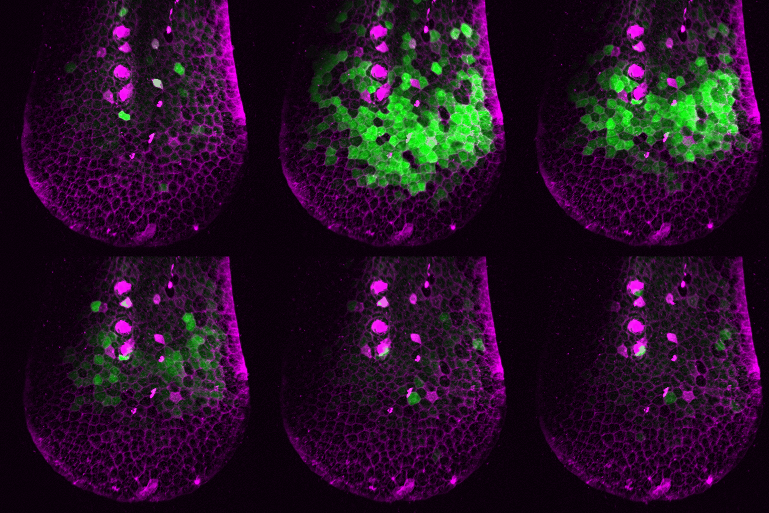
Confocal fluorescence microscopy
We design and build advanced confocal fluorescence microscopes under $150K. Key features:
-
3-4 lasers (free-space or fiber deliver)
-
Inverted microscope frame (Olympus IX71 or similar)
-
3D sample stage
-
High-speed laser scanning system (galvo-mirror-based)
-
3 PMT channels (customizable)
-
Software-based autofocusing
-
Automated image stitching and FOV registration
-
Advanced and unified image acquisition software (PyRPOC)
-
Can be integrated with advanced optical control systems (RPOC)
-
Longitudinal live cell imaging (additional device needed)

Confocal fluorescence microscope
The advanced confocal microscope is designed to support customized optical perturbation configurations, accommodating light sources ranging from UV to IR and from continuous-wave (CW) to femtosecond (fs) pulsed lasers.
The standard system includes three laser sources, three PMT detection channels, 3D sample positioning, and high-speed galvo scanning across a millimeter-scale field of view (FOV). The system can be integrated with stage-top incubators for long-term live-cell imaging over 72 hours without medium replacement (additional equipment required).
Image acquisition is facilitated by an advanced, unified software platform featuring autofocus, image stitching, field-of-view (FOV) registration, and fluorescence lifetime imaging microscopy (FLIM; additional TCSPC electronics and detectors required).